Digital Dental X-Rays in Dunedin, FL

See What a Visual Exam Can’t Show

Some dental problems develop where you cannot see them in a mirror — between two teeth, beneath an existing filling, or below the gumline. A visual examination is an important part of every dental visit, but it has limits. Digital dental X-rays give your dentist a second layer of information, so that what your dentist recommends is based on a more complete picture of what is actually happening in your mouth.

If you have ever wondered why X-rays are part of a routine dental exam, or whether the imaging is really necessary, those are fair questions. This page explains what digital dental X-rays show, what you can expect during the process, and why the imaging is used the way it is.

Schedule Your Dental Exam
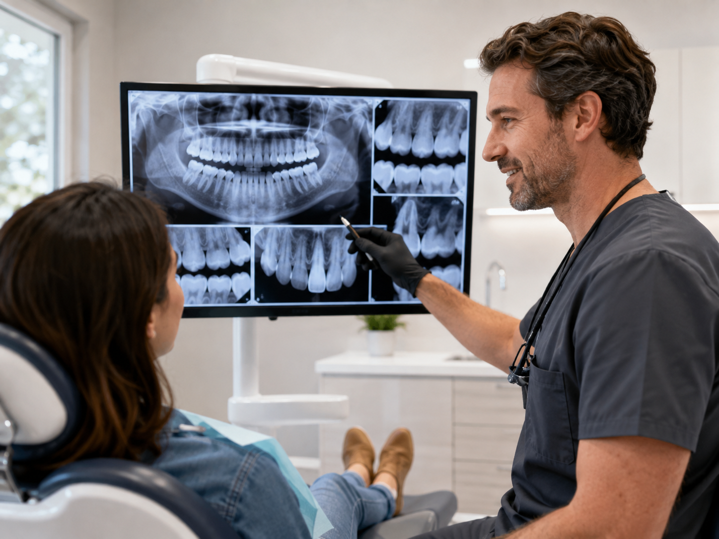
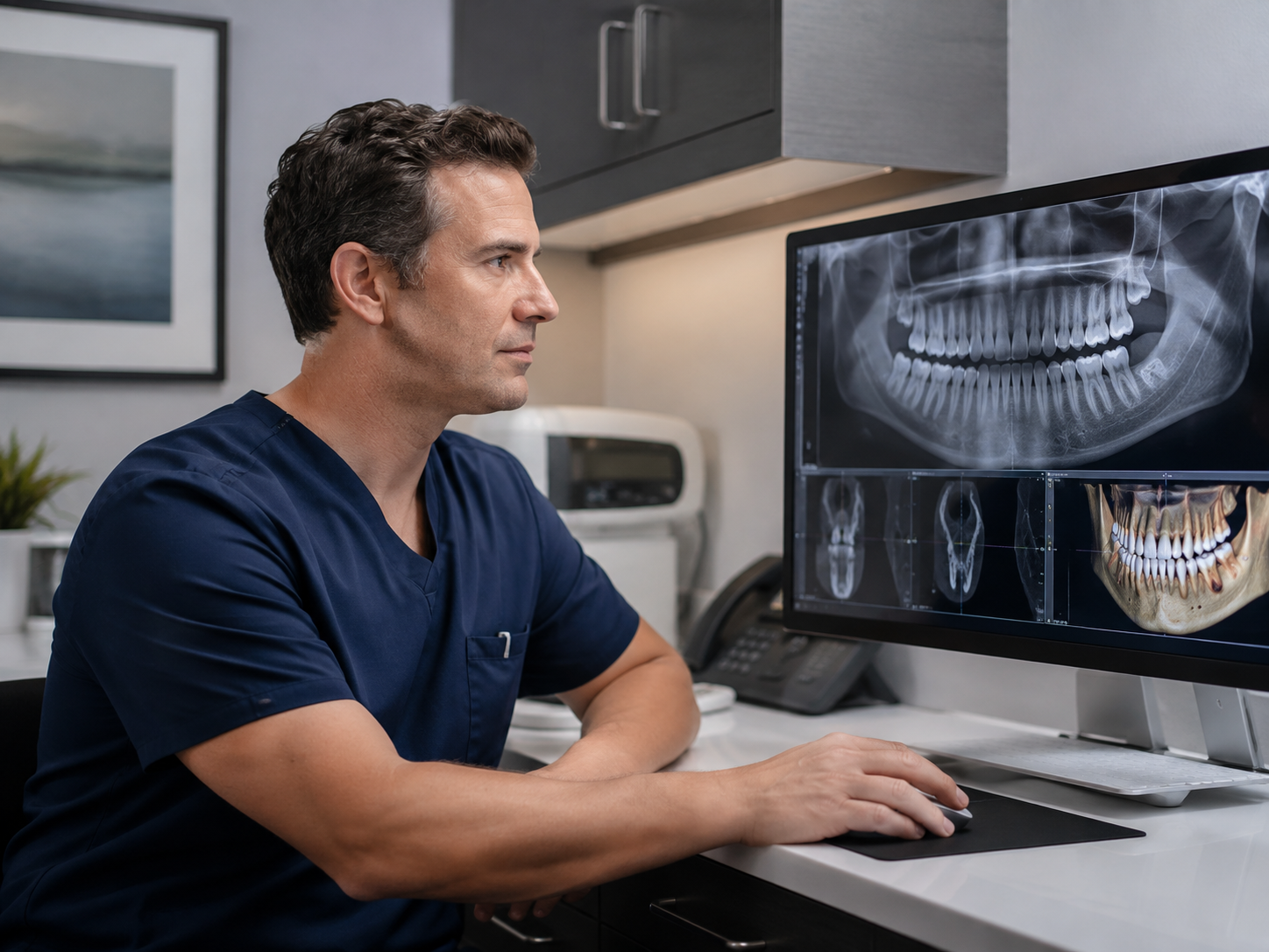
Dentist reviewing digital dental X-rays on a computer monitor during a dental exam
Seeing What a Visual Exam Can’t

Why Your Dentist Sometimes Needs to See More Than the Surface

A careful visual exam tells your dentist a great deal, but some dental conditions develop in places that simply cannot be evaluated by looking at the outside of a tooth. Digital dental X-rays help complete the picture.

What Digital X-Rays Can Help Your Dentist Evaluate

Some dental concerns may stay hidden during a routine visual exam alone. Depending on your situation, digital dental X-rays may help your dentist evaluate:

  • Decay developing between teeth
  • Bone loss below the gumline
  • Tooth roots and surrounding support structures
  • Changes around an existing crown or filling
  • Problems that may not yet be causing noticeable symptoms
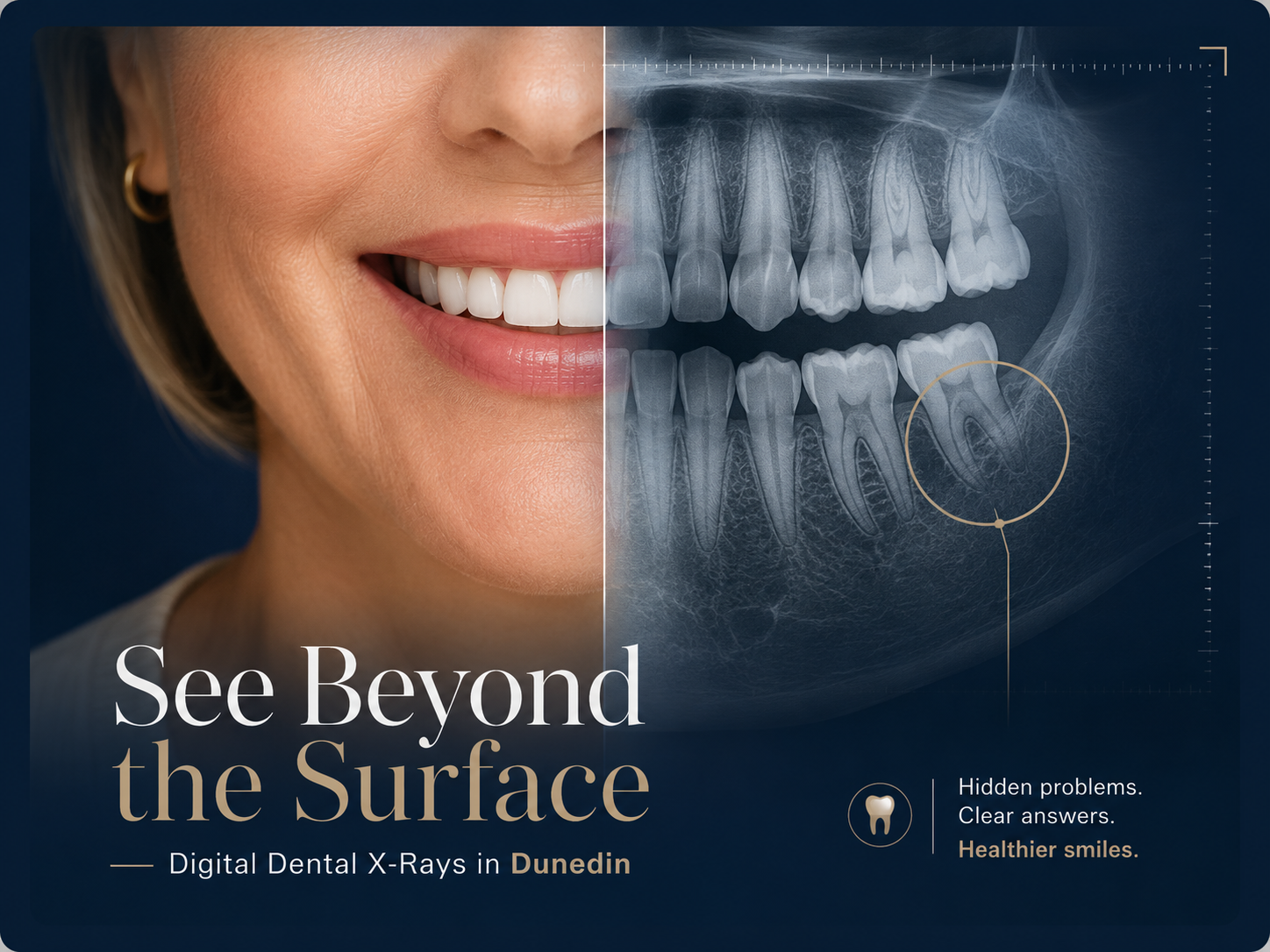
Digital dental X-rays showing hidden problems beneath the surface of a smile

Decay that forms between teeth is one example. So is bone loss that develops slowly below the gumline, or a problem developing around the root of an existing crown. By the time any of these cause noticeable symptoms, they may have been present for some time.

X-rays are not taken to find problems that do not exist. They are taken because some things cannot be assessed without them — and because catching a developing issue earlier is generally easier to address than waiting for it to become symptomatic. Digital dental X-rays extend what your dentist can evaluate during an exam so that the recommendation you receive is based on the fullest picture available.

What Are Digital Dental X-Rays?

Digital dental X-rays use an electronic sensor instead of traditional X-ray film, giving your dentist detailed images that can be viewed and reviewed on a computer within seconds.

How Digital X-Rays Capture the Image

Digital dental X-rays are a modern form of dental radiography that uses an electronic sensor instead of traditional X-ray film. When the sensor captures an image, it sends it directly to a computer screen — typically within seconds.

Your dentist can then view, enlarge, and examine the image alongside what was found during your clinical dental exam.
What the X-Ray Image Actually Shows

The basic imaging principle is similar to traditional dental X-rays. A small amount of radiation passes through the teeth and surrounding structures while different tissues absorb it at different levels.

Denser structures such as enamel and bone appear differently on the resulting image, helping your dentist evaluate areas that may not be visible during a visual examination alone.

Digital X-Rays vs. Traditional Film

What changes with digital radiography is not the basic purpose of the X-ray, but how the image is captured, processed, reviewed, and stored. Digital images become available quickly and can be enlarged directly on a monitor.

Modern digital systems also change the amount of radiation required compared with traditional film-based X-rays, while eliminating the need to chemically process traditional X-ray film.

How We Use Digital Imaging in Your Exam

At Dunedin Dental Associates, digital imaging is used as part of the comprehensive dental exam process when it is clinically indicated.

Your images can be reviewed at chairside, giving your dentist the opportunity to walk you through relevant findings and explain what they mean before discussing any recommended next step.

What Can a Dental X-Ray Show That an Exam Cannot?

This is one of the most practical questions a patient can ask. Dental X-rays do not replace a visual examination — they complement it. Depending on the type of image taken and your clinical situation, X-rays can help your dentist evaluate areas that simply cannot be seen by looking at the surface of your teeth.

01

Between the Teeth

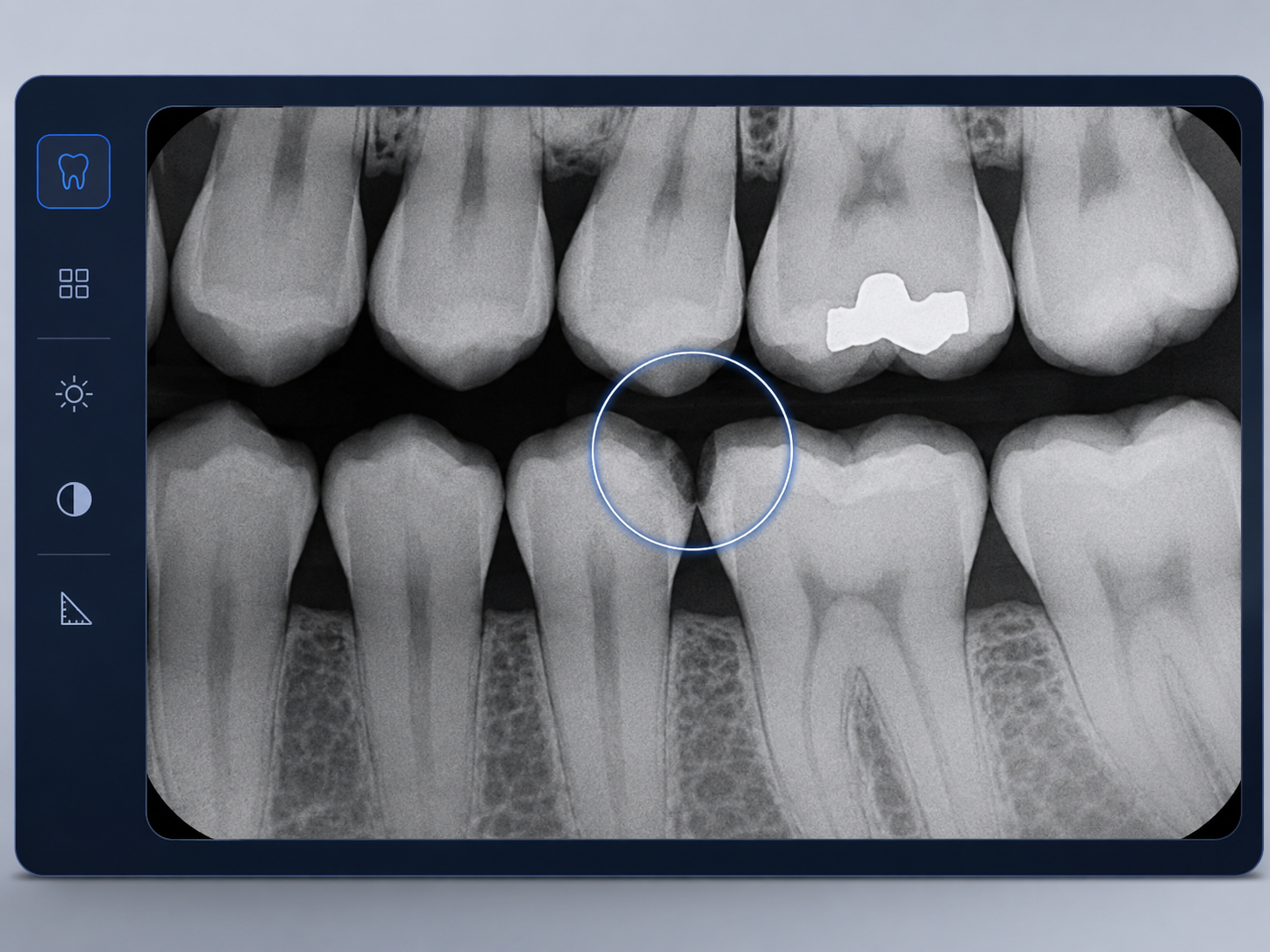
Decay can begin where two teeth touch — an area your dentist cannot always inspect directly during a visual exam. Digital dental X-rays can help reveal decay forming between teeth before it becomes obvious from the outside.

Imaging may also provide useful information about areas beneath or around existing fillings, crowns, and bridges when your dentist needs to evaluate what cannot be seen from the surface alone.

Bitewing digital dental X-ray showing possible decay between neighboring teeth
02

Below the Gumline

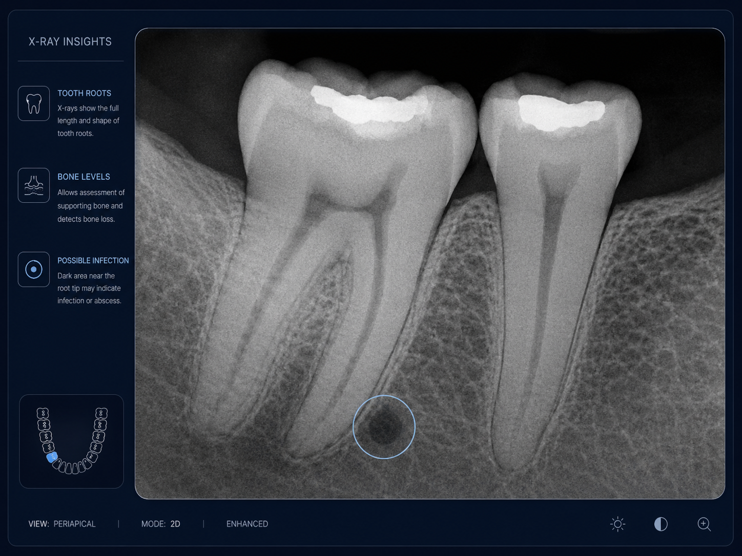
X-rays allow your dentist to evaluate the bone supporting your teeth and identify changes in bone levels that may be associated with periodontal disease.

They can also show the roots of your teeth and the surrounding structures, where certain infections or abscesses may develop. Problems in these areas can exist beneath the surface even when the visible part of the tooth appears relatively normal.

Periapical dental X-ray showing tooth roots, supporting bone levels, and a possible root-area infection
03

Beyond What Has Erupted

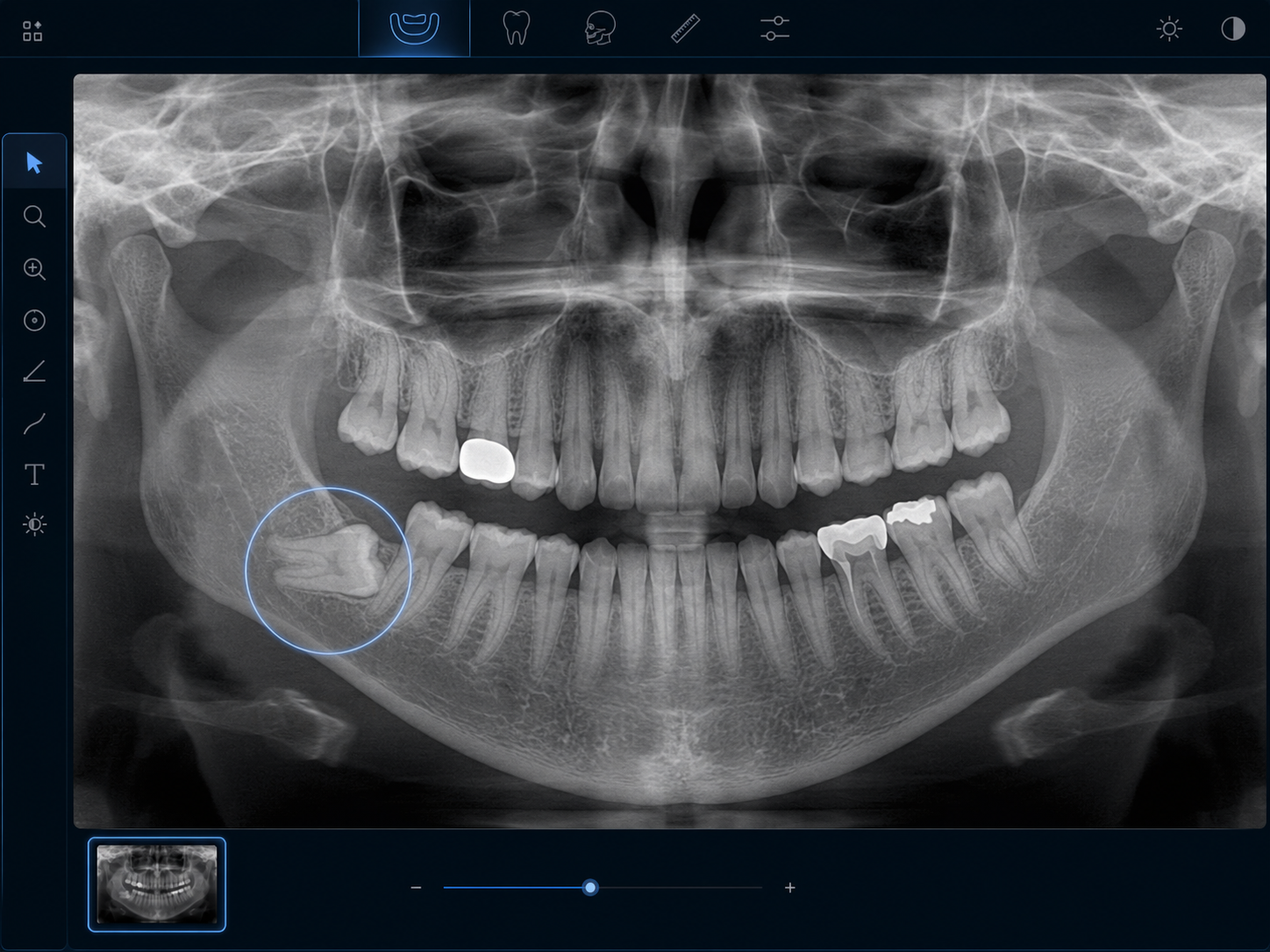
Some teeth remain beneath the gumline or do not erupt normally. Panoramic X-rays can help your dentist evaluate impacted or unerupted teeth along with broader areas of the jaw that cannot be assessed through a routine visual examination.

Depending on the image and clinical need, X-rays may also reveal certain bone changes or abnormalities and provide a baseline that can be compared with future images.

Panoramic dental X-ray showing an impacted tooth beneath the gumline

A Baseline Today Can Make Changes Easier to Recognize Tomorrow

Not every X-ray is taken because your dentist already suspects a specific problem. Baseline images can also provide a point of comparison over time. Changes in bone levels, developing decay, or other findings can then be compared with earlier images rather than being evaluated without that history.

X-rays that reveal infections around a tooth root are a good example of this diagnostic value. What feels like vague tooth pain or sensitivity may sometimes be connected to something happening below the visible surface. Learn more about tooth infections and abscesses and why a professional examination is necessary to determine the cause.

Digital X-Rays vs. Traditional Film X-Rays

Traditional film-based X-rays have been used in dentistry for many years, and they remain diagnostically useful. Digital radiography is a more modern approach that changes how images are captured, processed, reviewed, and stored. The difference is not only technical — it also changes what the experience is like for both the dentist and the patient.

Modern Digital Imaging

Digital X-Rays

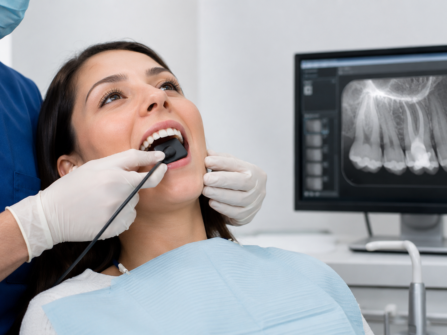
Digital dental X-rays use an electronic sensor instead of traditional film. Once the image is captured, it is transmitted directly to a computer, usually within seconds. This makes the imaging process faster and more efficient, while allowing your dentist to review the result during the same appointment.

Because the image is displayed digitally, it can also be enlarged and reviewed in greater detail as part of your dental exam.

Patient receiving a digital dental X-ray with an electronic sensor during a dental exam
Chairside Review

Faster Review and More Flexible Image Use

With digital radiography, the image is available almost immediately. Your dentist can review it with you at chairside, enlarge areas of interest, and adjust brightness or contrast after capture when that helps interpretation.

Digital files are also easier to store and retrieve over time. That matters when your dentist wants to compare current images with earlier ones to monitor changes in bone levels, watch a developing area, or evaluate how a condition has progressed.

Dentist reviewing digital dental X-rays with a patient on a chairside monitor
Earlier Film Workflow

Traditional Film X-Rays

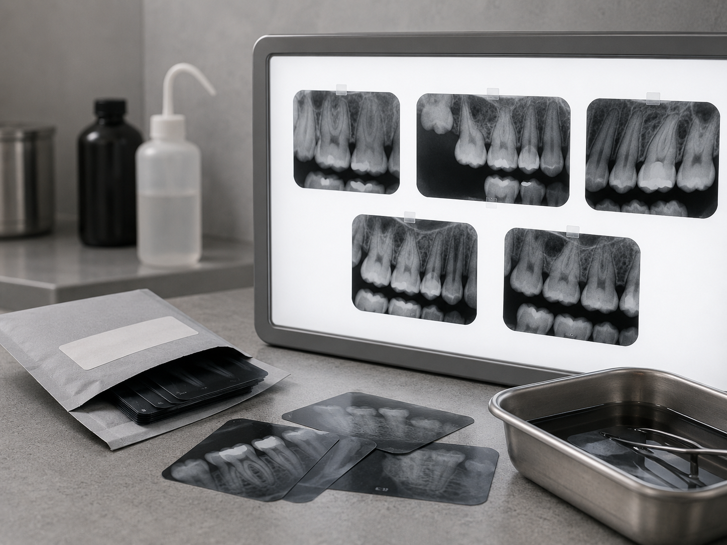
Traditional dental X-rays rely on physical film that is exposed in the mouth and then processed after the image is taken. The core diagnostic principle is the same, but the workflow is less immediate than modern digital systems.

Film-based X-rays must be developed before they can be reviewed, and once the image is processed, it cannot be adjusted the way a digital image can. Physical films are also stored manually rather than being saved as digital files.

Traditional dental X-ray films displayed on a light box with physical film packets nearby
Practical Comparison

The Difference in Daily Practice

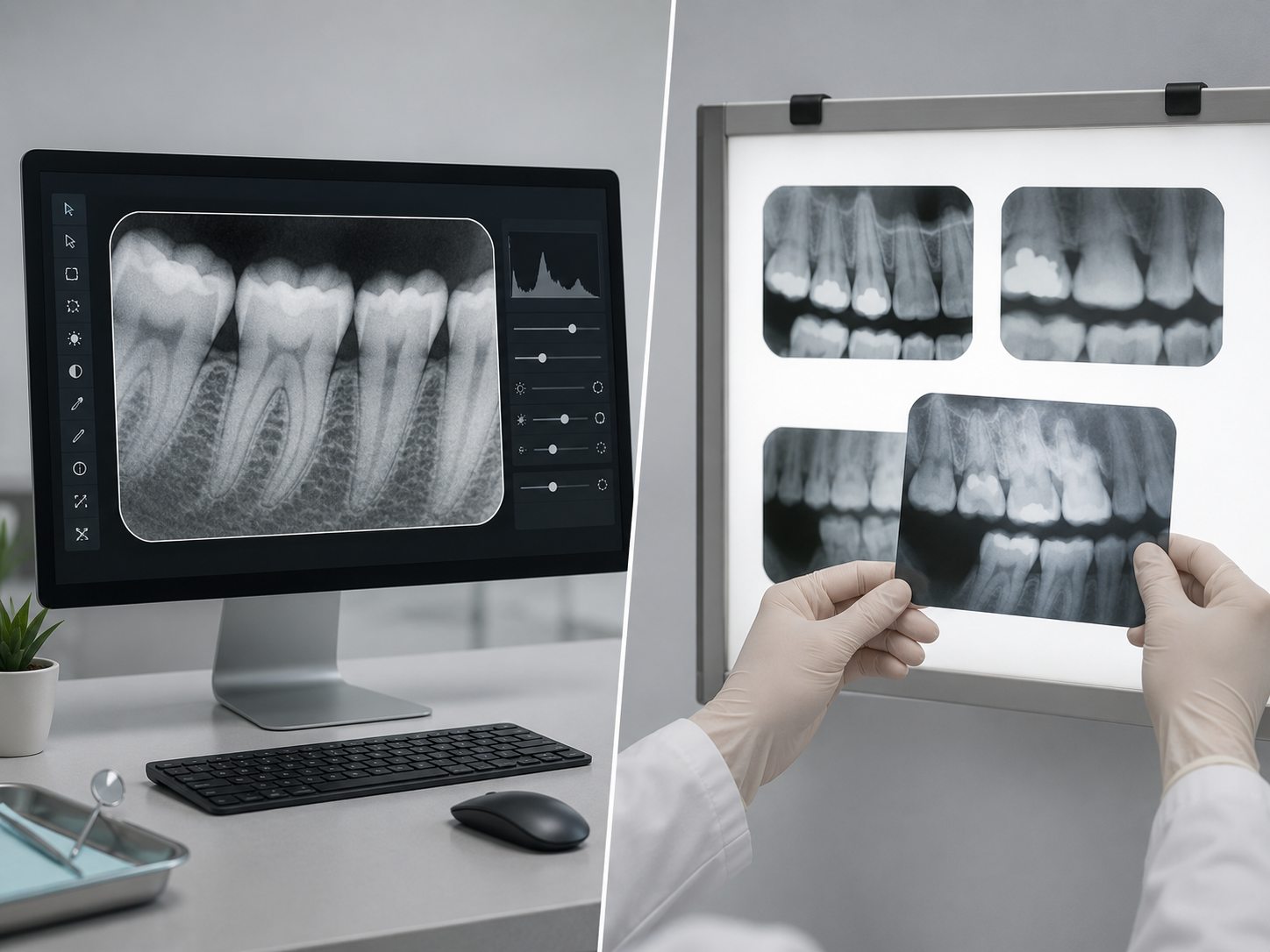
In practical terms, digital and traditional film X-rays differ in several ways. Digital systems use electronic image capture, provide images in seconds, allow chairside enlargement and review, and store records as digital files. Traditional film requires chemical development, physical film storage, and a separate viewing method.

Digital X-rays also generally require less radiation than conventional film-based systems and eliminate the chemical processing waste associated with traditional film. These advantages are part of why digital radiography has become the standard across modern dental practices.

Side-by-side comparison of digital dental X-rays on a computer and traditional film X-rays on a light box

Why Most Modern Dental Practices Use Digital Radiography

The transition from traditional film-based X-rays to digital systems has become standard across modern dentistry for practical reasons. Digital dental X-rays support faster image review, streamlined storage, chairside discussion, and reduced radiation exposure compared with conventional film. At Dunedin Dental Associates, digital imaging is used when clinically indicated so that your dentist can make recommendations based on a clearer and more complete view.

Digital X-Ray Safety in Dunedin

How Much Radiation Do Digital Dental X-Rays Use?

Radiation exposure is one of the most common questions patients ask about dental X-rays — and it is a reasonable question. Modern digital dental X-rays use digital receptors that can reduce exposure compared with conventional film-based imaging.

The goal is not to take as many images as possible. It is to obtain the diagnostic information your dentist actually needs while keeping exposure as low as reasonably achievable.

01

Digital Image Capture

Digital receptors are more efficient than conventional film, allowing the dental team to obtain diagnostic images while reducing radiation exposure compared with traditional film-based systems.

02

Taken for a Clinical Reason

Dental X-rays should be selected according to the information your dentist needs — not simply taken on the same schedule for every patient regardless of symptoms, risk, or previous images.

03

Your Situation Guides the Decision

Age, oral health, symptoms, disease risk, previous imaging, pregnancy, and the type of information needed can all influence which images — if any — your dentist recommends.

Patient-Specific Radiation Safety

Keeping Exposure as Low as Reasonably Achievable

Any medical use of X-rays involves balancing diagnostic benefit with radiation exposure. Dental professionals use the ALARA principle — “as low as reasonably achievable” — so that necessary images provide useful clinical information without unnecessary exposure.

Current dental guidance emphasizes choosing X-rays according to individual clinical need, using digital receptors, limiting the X-ray beam to the area that needs to be examined, and making use of suitable previous images whenever possible.

Radiation from dental imaging represents a small contribution to a person's overall exposure from natural and medical sources. Your dentist still considers whether the expected diagnostic information justifies taking each image.

If you have questions about pregnancy, your child's X-ray frequency, previous imaging, or radiation exposure in general, tell your dentist before imaging begins. The goal is to explain why an X-ray is being recommended and how it relates to your individual dental care.

i

Current ADA guidance does not recommend routine lead aprons or thyroid collars for dental imaging. Modern equipment, appropriate image selection, digital receptors, and beam restriction are considered more effective approaches to minimizing unnecessary exposure. Applicable state and local radiation regulations are still followed.

Digital Receptor Focused Field Clinical Need
Digital Efficient Image Capture
Selective Patient-Specific Imaging
Explained Clear Clinical Reason

Lorem ipsum dolor sit amet, consectetur adipiscing elit. Ut elit tellus, luctus nec ullamcorper mattis, pulvinar dapibus leo.

TYPES OF DIGITAL DENTAL X-RAYS

Different X-Rays Answer Different Dental Questions

Not every dental X-ray shows the same thing. Your dentist chooses the type of image based on what needs to be evaluated — from decay between teeth to the roots, supporting bone, or the broader structure of your jaws.

Bitewing X-Rays

Bitewing X-rays show the upper and lower back teeth together in a single image. You bite gently on a small tab or digital sensor holder while the image is taken.

These X-rays are especially useful for detecting decay between teeth and evaluating the bone levels that support them. They are commonly used during routine dental exams when your dentist needs a closer look at areas that cannot be seen directly.

Periapical X-Rays

When your dentist needs to evaluate one particular tooth more closely, a periapical X-ray can show the entire tooth — from the crown above the gumline to the tip of the root — along with the surrounding bone.


It may be recommended when investigating tooth pain, examining the root area for signs of infection, or evaluating changes in the bone around a specific tooth.

Panoramic X-Rays

A panoramic X-ray provides a broad view of your entire mouth in one image, including the teeth, jaws, sinuses, and surrounding structures. Instead of placing a sensor inside your mouth, the imaging unit rotates around your head.

Panoramic imaging is often useful when evaluating wisdom teeth, jaw structure, impacted teeth, or planning certain procedures such as dental implants. It provides different information from bitewing and periapical X-rays rather than replacing them.

Your Dentist Chooses the Image You Actually Need

You do not need every type of X-ray at every dental visit. The appropriate image depends on what your dentist is trying to understand, your current oral health, symptoms, previous X-rays, and your individual risk factors.

The goal is simple: obtain the right diagnostic information for your situation, rather than taking images that are not clinically necessary.

Your Digital Imaging Experience

Digital Imaging Should Give You Answers — Not More Questions

Dental X-rays are most useful when they are part of a conversation, not just a procedure. At Dunedin Dental Associates, digital images are reviewed with you so you can understand what your dentist sees, what it means, and whether anything needs attention.

Step 01

Images Are Taken for a Clinical Reason

Digital X-rays are taken when your dentist needs information that cannot be gathered from the visual exam alone.

The goal is not simply to collect images. It is to answer a specific clinical question and give your dentist a more complete picture of your oral health.

Step 02

Your Dentist Reviews the Images

Dunedin Dental Associates uses Planmeca digital imaging technology as part of the comprehensive dental exam process when imaging is clinically indicated.

Once captured, the digital images can be viewed at chairside and evaluated alongside what your dentist found during the examination.

Step 03

You See What Your Dentist Sees

Digital images can be enlarged and adjusted when closer review is helpful. Previous imaging may also be compared with current images to look for meaningful changes over time.

Instead of hearing only that something was found, you can see the area your dentist is discussing and understand why it matters.

Step 04

The Findings Are Explained Clearly

An X-ray does not automatically mean treatment is needed. Sometimes an image confirms that nothing unusual is visible. In other situations, it may reveal a small area that should simply be watched.

Your dentist explains the findings in plain language before making any recommendation.

Step 05

You Leave Knowing What Comes Next

By combining your digital X-rays with the clinical exam, your dentist can explain what appears healthy, what may need monitoring, and what — if anything — should be addressed.

For a broader look at how imaging fits into your visit, see our comprehensive dental exam process.

Clearer Information Leads to Better-Informed Decisions The goal is not to overwhelm you with images or technical language. It is to help you understand your oral health, see the information behind your dentist’s recommendation, and feel confident about the next step.
Your Digital X-Ray Appointment

What to Expect During Your Digital X-Ray Appointment

Digital dental X-rays are a routine part of the exam process when clinically appropriate. The process is usually quick: the images are captured, reviewed with your examination findings, and then explained so you know exactly what comes next.

Your Dentist Reviews the Images

The images appear on the screen almost immediately. Your dentist reviews them together with the visual examination, looking at areas such as bone levels, spaces between teeth, roots, and other clinically relevant structures.

You Discuss What Comes Next

Your dentist explains what the images show and what that means for your oral health. If something needs attention, you will understand why. If the images look healthy, that is useful information too.

Swipe horizontally to view each step
Appointment Images Are Taken Digital image capture Dentist Reviews Exam + imaging together Findings Discussed Clear next step
What happens Sensor positioned and image captured Images evaluated with your clinical exam Findings explained in plain language
Typical timing Seconds per image Review begins almost immediately Discussed during the same visit
What is evaluated The specific area your dentist needs to see Bone, tooth surfaces, roots, and other relevant structures Healthy findings, areas to monitor, or treatment needs
Patient experience Quick sensor positioning Images visible on the computer Opportunity to ask questions
Digital record Captured digitally Stored with your record Available for future comparison
Main purpose Gather information Understand findings Make an informed decision

A Quick Process That Becomes Part of Your Dental Record

Digital X-rays generally add only a short amount of time to the appointment. Once captured, the images are stored as digital files in your dental record and can be compared with future imaging when your dentist needs to evaluate changes over time.

How Often Do You Need Dental X-Rays?

There is no single X-ray schedule that is right for every patient. The timing depends on your oral health, your history, and what your dentist needs to evaluate during the exam.

The American Dental Association recommends that dental X-rays be selected according to individual clinical circumstances rather than taken automatically at the same interval for everyone.

Your X-Ray Schedule Should Be Based on Your Needs

Several factors can influence how often your dentist recommends digital dental X-rays, including your age, current oral health, cavity risk, periodontal condition, symptoms, previous treatment, and the findings from earlier images.

If you have a history of tooth decay, active gum disease, certain restorations, or a new dental concern, your dentist may recommend imaging more frequently so changes can be evaluated appropriately.

On the other hand, if your teeth and gums have remained stable, previous X-rays have shown no developing concerns, and you have no new symptoms, you may need imaging less often.

The goal is not to take X-rays simply because a certain amount of time has passed. The goal is to take the right image when it provides useful information for your care.

If you have recently had dental X-rays taken at another office, bring or send those records to Dunedin Dental Associates when possible. Reviewing recent images may help your dentist avoid unnecessary repeat imaging while still having the information needed to evaluate your oral health.

Common Questions About Digital Dental X-Rays

Digital dental X-rays use a very small amount of radiation. According to the American Dental Association, the radiation from a full set of digital dental X-rays is comparable to a fraction of the background radiation that people are exposed to through everyday natural sources. Protective equipment — including a lead apron and thyroid collar — is used during every X-ray appointment. Your dentist will only recommend imaging when it is clinically indicated.

Yes. Digital X-ray technology generally requires less radiation than conventional film-based X-rays to produce a comparable image. This is one of the reasons digital systems have become the standard in modern dental practices.

Not necessarily. X-ray frequency is determined by your individual clinical circumstances — including your oral health history, current findings, and decay risk — not by a fixed schedule. Your dentist will recommend imaging when it is clinically appropriate for your situation.

Yes. X-rays are particularly useful for detecting decay that forms between teeth, where it is not visible during a visual examination. Catching decay earlier typically makes it easier to treat and less likely to require more extensive work. That said, X-rays are interpreted alongside the clinical exam — no single diagnostic tool captures everything on its own.

No. The imaging itself is painless. The sensor placed inside the mouth can sometimes feel slightly uncomfortable depending on the area being imaged, but the process is typically quick. If you find the process difficult — for example, if you have a sensitive gag reflex — let your dental team know before the appointment begins.

The main differences are in how the image is captured and processed. Digital X-rays use an electronic sensor that transmits the image to a computer screen within seconds, require less radiation than traditional film, and allow the dentist to review and enlarge the image at chairside. Traditional film requires chemical processing and is stored as a physical negative.

This is a question best answered in conversation with your dentist and your obstetrician. In general, dental X-rays during pregnancy are considered low-risk with appropriate shielding, and the American Dental Association supports providing necessary dental care during pregnancy. However, individual circumstances vary. Let your dental team know if you are pregnant or think you may be.

Dental X-rays — particularly panoramic images — can provide useful information about jaw bone and surrounding structures that may inform discussions about dental implant planning. In some cases, a more advanced three-dimensional CBCT scan may be recommended specifically for implant evaluation. Your dentist will advise on the appropriate imaging based on your specific situation.

Clear Answers Before the Next Step

Clearer Information Leads to Better Dental Decisions

You should not have to make dental decisions based on incomplete information. When your dentist can evaluate both what is visible during the exam and what the imaging shows, the recommendation you receive reflects a more complete picture of your oral health.

Digital dental X-rays do not guarantee that every dental problem will be found. They do, however, extend what your dentist can assess beyond the visible surface — while providing images that can be reviewed with you before any treatment decision is made.

Start With an Exam — Not a Treatment Decision

If you are due for a dental exam or have a question about a specific tooth, our team at Dunedin Dental Associates can evaluate what is happening, explain what the images show, and help you understand what — if anything — should happen next.

Directions
Office Hours
Monday | 8am – 5pm
Tuesday | 8am – 5pm
Wednesday | 8am – 5pm
Thursday | 8am – 5pm
Friday | By Appointments Only
Saturday | Closed
Sunday | Closed

Copyright © 2026